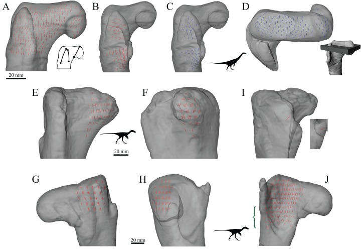
Figure 21. The main architectural features of cancellous bone in the proximal femur of Falcarius utahensis and Troodontidae sp.
(A) Vector field of u1 in the proximal femur of Falcarius (UMNH VP 12361), viewed as a 3D slice parallel to the coronal plane and through the middle of the bone. Schematic inset illustrates three main trajectories in this image, which are not too dissimilar from the patterns observed in humans and large non-avian theropods (cf. Figs 15, 19). (B, C) Vector field of u1 (B) and u2 (C) in the lesser trochanter of Falcarius, in oblique anterolateral view. (D) Vector field of u2 in the proximal femur of Falcarius, in a 3D slice parallel to the axial plane and through the femoral head. Main image is shown in axial view (anterior is toward bottom of page), with inset illustrating the region illustrated in context of the whole bone. (E, F) Vector field of u1 in the femoral head and inferior neck of Troodontidae sp. (MOR 748) in anterior (E) and medial (F) views. (G, H) Vector field of u1 in the region of the greater trochanter of Troodontidae sp. (MOR 553s-7.28.91.239) in posterior (G) and lateral (H) views. (I) Orientation of u1 in the lesser trochanter, or immediate region thereof, of Troodontidae sp., in oblique anterolateral view (main image illustrates MOR 748; inset illustrates MOR 553s-7.28.91.239). (J) Vector field of u1 throughout the metaphysis of Troodontidae sp. (MOR 553s-7.28.91.239), illustrating increasing obliquity and disorganization of vectors in the distal metaphysis and transition to the diaphysis (region with braces).

