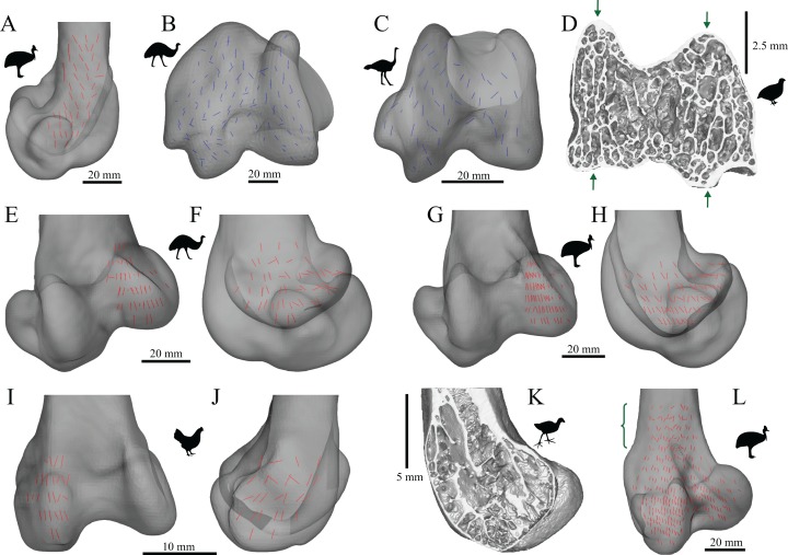
Figure 24. The main architectural features of cancellous bone in the distal femur of birds.
(A) Vector field of u1 in the central metaphysis of a southern cassowary, Casuarius casuarius (QMO 30105), in a 3D slice, parallel to the sagittal plane and between the condyles, shown in lateral view. Note the weakly developed double arcuate pattern. (B, C) Vector field of u2 in a 3D slice through the middle of the condyles in an ostrich, Struthio camelus (MV R.2385, B), and a emu, Dromaius novaehollandiae (QMO 11685, C), shown in distal view. Note the ‘butterfly pattern’ in both examples. (D) Isosurface rendering of cancellous bone in the distal condyles of a Japanese quail, Coturnix japonica (PJB coll.), sectioned in the axial plane; notice the ‘butterfly pattern’ between the arrows. (E–H) Vector field of u1 in the medial condyle of Dromaius novaehollandiae (QMO 16140, E, F) and Casuarius casuarius (QMO 30604, G, H), shown in anterior (E, G) and medial (F, H) views. (I, J) Vector field of u1 in the lateral condyle of a chicken, Gallus gallus (PJB coll.), shown in anterior (I) and lateral (J) views. (K) Isosurface rendering of cancellous bone in the medial condyle of a purple swamphen, Porphyrio porphyrio (PJB coll.), sectioned in the sagittal plane. (L) Vector field of u1 throughout the entire distal femur of Casuarius casuarius (QMO 31137), illustrating increasing obliquity and disorganization of vectors in the proximal metaphysis and transition to the diaphysis (region with braces).

