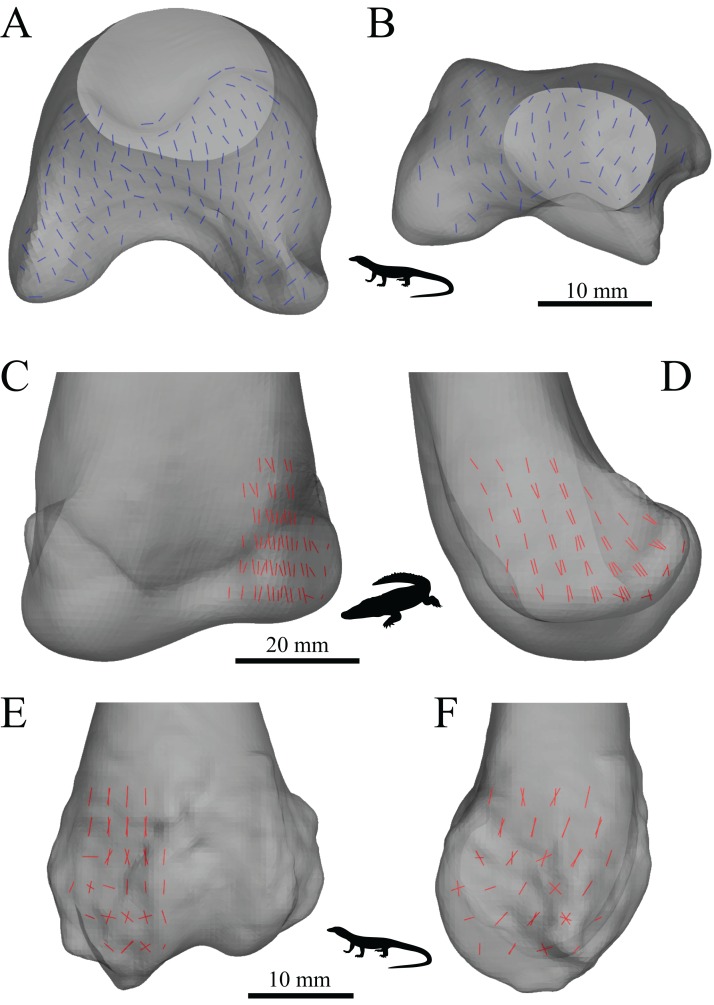
Figure 25. The main architectural features of cancellous bone in the distal femur of extant sprawling reptiles.
(A, B) Vector field of u2 in a 3D slice through the middle of the condyles in a freshwater crocodile, Crocodylus porosus (QMJ 48127, A), and a Komodo dragon, Varanus komodoensis (AM R.106933, B), shown in proximal view. (C, D) Vector field of u1 in the medial condyle of Crocodylus porosus (QMJ 48127), shown in anterior (C) and medial (D) views. (E, F) Vector field of u1 in the lateral condyle of a Spencer’s goanna, Varanus spenceri (QMJ 484416), shown in anterior (E) and lateral (F) views.

