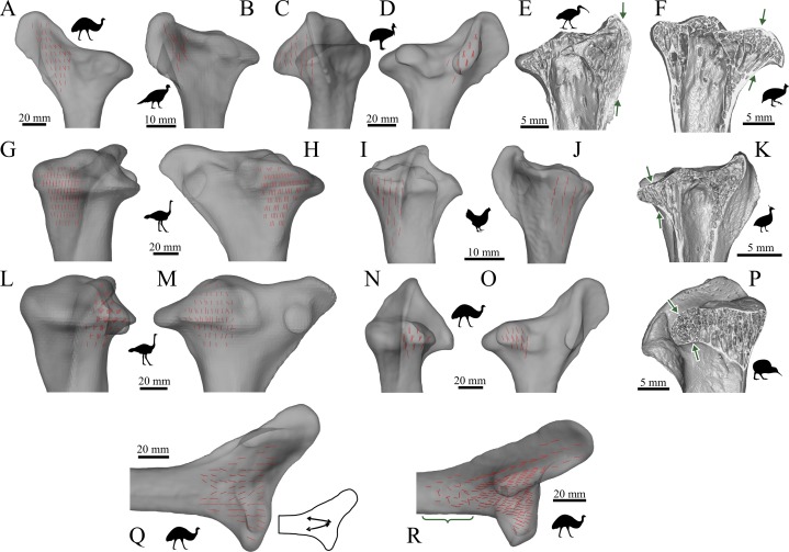
Figure 31. The main architectural features of cancellous bone in the proximal tibia of birds.
(A, B) Vector field of u1 in the anterior (cranial) cnemial crest of an emu, Dromaius novaehollandiae (QMO 11686, A), and turkey, Meleagris gallopavo (RVC turkey 1, B), shown in medial view. (C, D) Vector field of u1 in the lateral cnemial crest of a southern cassowary, Casuarius casuarius (QMO 30105), shown in anterior (C) and lateral (D) views. (E) Isosurface rendering of cancellous bone in the anterior cnemial crest of an Australian white ibis, Threskiornis moluccus (PJB coll., between arrows), sectioned in the sagittal plane. (F) Isosurface rendering of cancellous bone in the lateral cnemial crest of a guineafowl, Numida meleagris (PJB coll., between arrows), sectioned in the plane of the crest. (G–J) Vector field of u1 under the medial condyle of an ostrich, Struthio camelus (MV R.2385, G, H), and chicken, Gallus gallus (PJB coll., I, J), shown in posterior (G, I) and medial (H, J) views. (K) Isosurface rendering of cancellous bone under the medial condyle of an elegant-crested tinamou, Eudromia elegans (UMZC 404.e, between arrows), sectioned in the sagittal plane. (L–O) Vector field of u1 under the lateral condyle of Struthio camelus (MV R.2711, L, M) and Dromaius novaehollandiae (QMO 11686, N, O), shown in posterior (L, N) and lateral (M, O) views. (P) Isosurface rendering of cancellous bone under the lateral condyle of a little spotted kiwi, Apteryx owenii (UMZC 378.iii, between arrows), sectioned in the coronal plane. (Q) Vector field of u1 in a 3D slice through the middle of the proximal metaphysis, cnemial crests and condyles of Dromaius novaehollandiae (QMO 11686), parallel to the sagittal plane. Schematic inset illustrates the moderately developed double-arcuate pattern present. (R) Vector field of u1 throughout the entire proximal tibia of Dromaius novaehollandiae (QMO 11686), illustrating increasing obliquity and disorganization of vectors in the distal metaphysis and transition to the diaphysis (region with braces).

