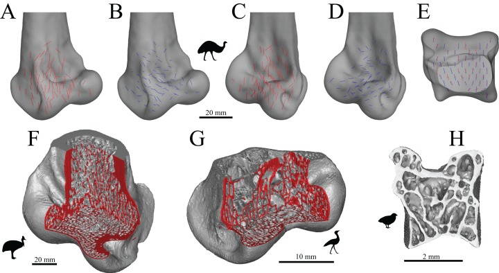
Figure 36. The main architectural features of cancellous bone in the distal tibiotarsus of birds.
(A–D) Vector field of u1 (A, C) and u2 (B, D) in the distal tibiotarsus of an emu, Dromaius novaehollandiae (QMO 16140), in oblique anterolateral (A, B) and oblique anteromedial (C, D) views. (E) Vector field of u1 (red) and u2 (blue) in the condyles of Dromaius novaehollandiae (QMO 16140) in proximal view (anterior is toward top of page). Note how both u1 and u2 are strongly aligned parallel to the sagittal plane. This particular specimen exemplifies a very stereotypical pattern that is observed in all large birds; the general pattern illustrated here was also observed in smaller species for which only limited fabric analysis was possible. (F) Isosurface rendering of cancellous bone in the distal tibiotarsus of a southern cassowary, Casuarius casuarius (QMO 30105), shown in oblique anteromedial view, with multiple cuts through the bone to illustrate 3D architecture. (G) Isosurface rendering of cancellous bone in the distal tibiotarsus of a bustard, Ardeotis australis (MVB 20408), shown in oblique anterolateral view, with multiple cuts through the bone to illustrate 3D architecture. (H) Isosurface rendering of cancellous bone in the distal tibiotarsus of a painted quail, Coturnix chinensis (PJB coll.), sectioned in the axial plane through the middle of the condyles and shown in proximal view (anterior is toward top of page). In (F and G), cut surfaces are coloured red to better show the nature of the cancellous bone architecture, in particular, the plate-like nature of many of the trabeculae, largely aligned parallel to the sagittal plane.

