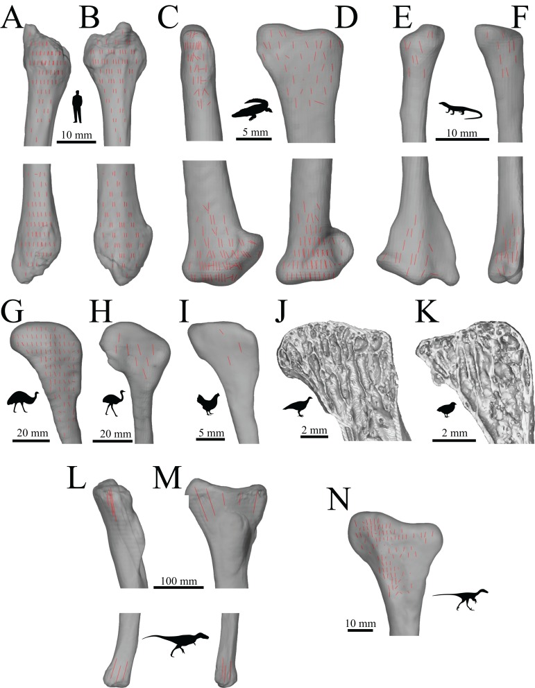
Figure 40. The main architectural features of cancellous bone in the fibula.
(A, B) Vector field of u1 in the human fibula, in anterior (A) and lateral (B) views. (C, D) Vector field of u1 in a freshwater crocodile, Crocodylus johnstoni (QMJ 47916), in anterior (C) and lateral (D) views. (E, F) Vector field of u1 in an Argus monitor, Varanus panoptes (QMJ 91981), in anterior (E) and lateral (F) views. (G–I) Vector field of u1 in the fibular head of an emu, Dromaius novaehollandiae (QMO 11686, G), greater rhea, Rhea americana (QMO 23517, H), and chicken, Gallus gallus (PJB coll., I), in lateral view. (J, K) Isosurface rendering of cancellous bone in the proximal fibula of a malleefowl, Leipoa ocellata (MVB 20194, J), and painted quail, Coturnix chinensis (PJB coll., K), sectioned in the plane of the head and shown in lateral view. (L, M) The dominant architectural direction of cancellous bone in the fibula of Allosaurus and tyrannosaurids, shown in anterior (L) and lateral (M) views. This is illustrated here with a 3D geometric model of the observed architecture, mapped to the fibula of Daspletosaurus torosus (TMP 2001.036.0001). (N) Vector field of u1 in the proximal fibula of Troodontidae sp. (MOR 553s-8.17.92.265), in lateral view.

