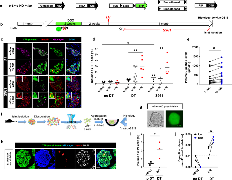
Figure 5. Smoothened (Smo) inactivation in α-cells facilitates their engagement into insulin production.
(a) Transgenes required for simultaneous α-cell lineage tracing, Smo co-receptor downregulation and DT-induced β-cell ablation. (b) Experimental design. (c) Smo inactivation in α-cells leads to insulin production when combined with β-cell loss (upper panels) or insulin receptor antagonism (bottom panels). IF were performed at on 3–5 consecutive sections per animal with similar results. The experiment was performed once, with mice treated asynchronously according to their availability. (d) Fraction of YFP+ cells producing insulin upon inactivation of Smo in α-cells combined with DT or S961. n=4,4,3 mice for Smowt/wt, Smofl/fl and Smofl/fl noDT, respectively; n=4,10,5 mice for Smowt/wt, Smofl/fl and Smofl/fl DT, respectively; n=4,5,6 mice for Smowt/wt, Smofl/fl and Smofl/fl S961, respectively. Center indicates the mean. Two-tailed Mann Whitney test, P=0.0079 Smofl/fl DT vs Smowt/wt DT, P=0.0095 Smofl/fl DT vs Smowt/wt S961 and P=0.0286 Smowt/wt vs Smowt/wt S961. Scale bars: 10 μm. (e) In vivo glucose challenge in α-Smo-KO mice. n=18 mice, P=0.012, Wilcoxon test, two-tailed. (f) Pipeline for α-cell sorting, in vitro pseudoislet reconstruction and functional tests. (g) Live imaging of 7-day-cultured pseudoislet reconstituted using α-cells from α-Smo-KO mice. Representative images from 3 independent experiments. (h) α-Smo-KO pseudoislet at day 7 of aggregation culture (immunofluorescence). Representative images from 3 independent experiments. Scale bar: 25 μm. (i) Percentage of YFP+ cells producing insulin in pseudoislets from control α-Smo-WT animals (no β-cell ablation) or α-Smo-KO mice after cell ablation (DT). n = 3 independent cohorts from 18 α-Smo-WT mice, and 3 independent cohorts from 18 α-Smo-KO mice, P=0.049, unpaired t-test, two-tailed. Center indicates the mean. (j) Glucose-stimulated C-peptide secretion. α-Smo-KO cells secrete C-peptide in response to glucose in vitro, while α-Smo-WT (no DT control) cells have no measurable secretion. n = 3 independent cohorts obtained from 18 α-Smo-Wt mice, n=3 independent cohorts obtained from 18 α-Smo-KO mice, P=0.048, paired t test, one-tailed. The dash line indicates the detection threshold documented by manufacturer (0.0127 nmole/pseudoislet/hour). All values are means ± s.e.m. See Supplementary Tables 1f-l for source data.

