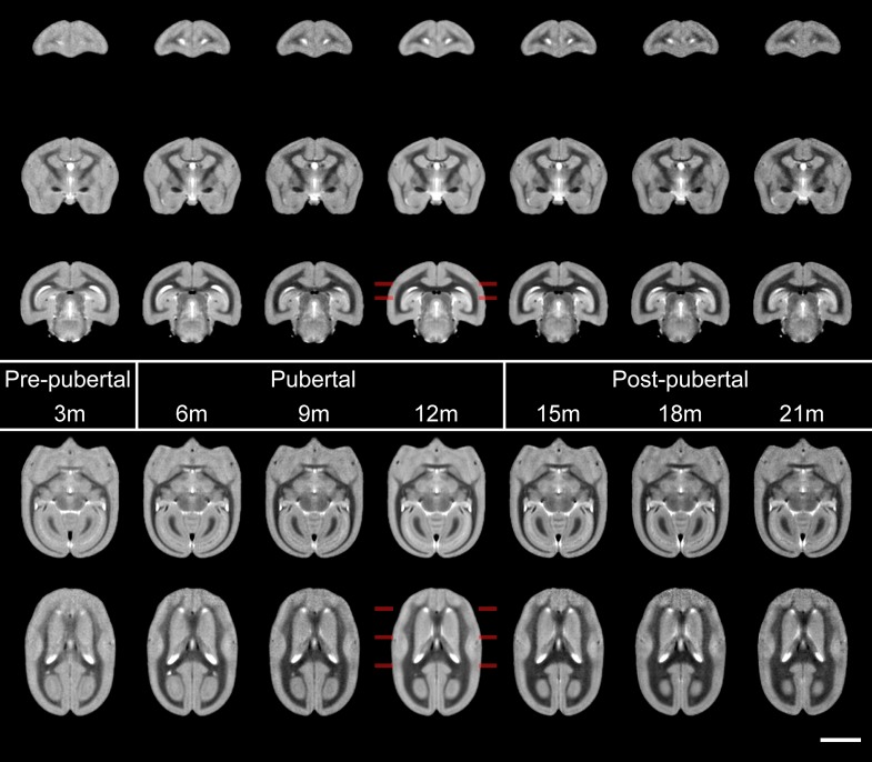
Figure 4.
Sections from mean templates of animals produced at regular intervals from 3 to 21 months, from prepuberty, through puberty and postpubertal stages from left to right. With the imaging parameters chosen, WM yields the darkest intensity, followed by GM and then CSF yields the brightest. Changes in WM are apparent at each age point: not only does it appear markedly thicker, but darker with time. Visually it appears to increase in volume at the expense of surrounding GM. Marked increases in white matter include that within the prefrontal cortex (coronal sections: top row), internal capsule (middle row 2) and corpus callosum (bottom row) and the anterior commissure (horizontal sessions: top row) and visual cortices (bottom row). Changes with age are readily appreciated visually with the simulated development movie (Supplementary Movie 1). Coronal sections are taken at 14, 6, and -2 mm from the intra-aural line, horizontal sections at 0 and 3.5 mm from the AC–PC line, indicated at the 12 m template. Scale bar is 5 mm.

