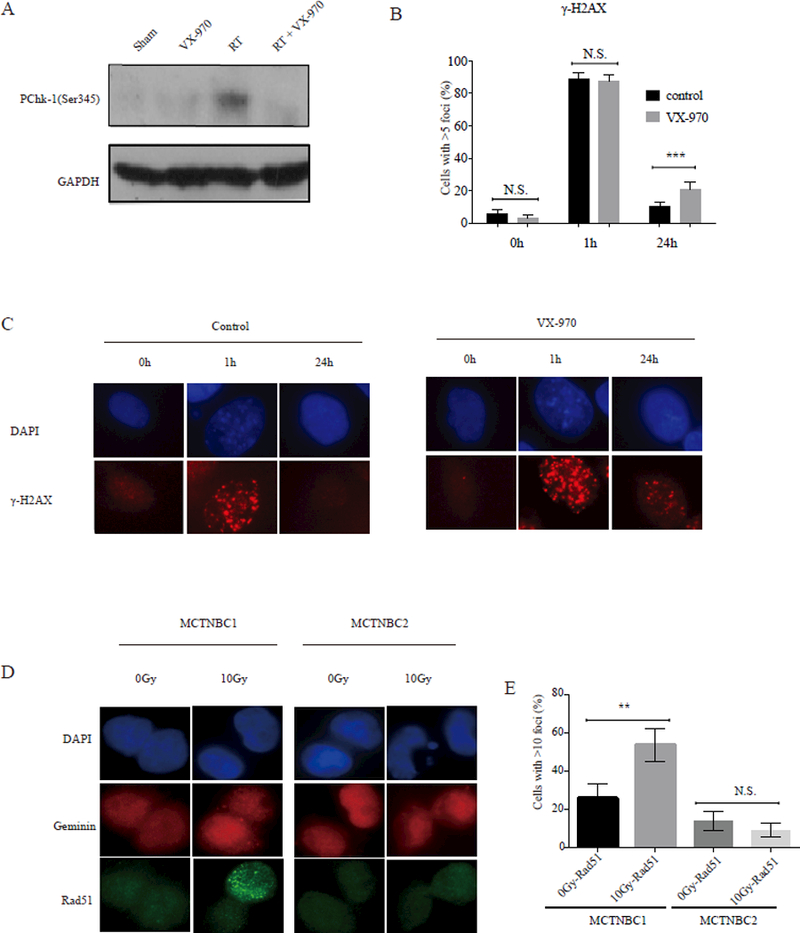
Figure 6.

DNA damage signaling and repair in TNBC PDXs treated with VX-970, RT, or the combination (A) Western blotting analysis of phosphorylated Chk-1 (pChk-1) following treatment with either sham RT, RT (2 Gy x 5 days), VX970 (60 mg/kg daily for five days) or RT + VX-970 administered one hour prior to daily fractionated RT administered over five days, as shown in the schema in figure 5A. MCTNBC1 tumor samples were harvested four hours after the last radiation treatment on day five. Samples were pooled from three mice per group. (B, C) TNBC PDX cells were pretreated, ex-vivo, with vehicle (DMSO) or VX-970 (80 nM) for one hour prior to exposure to 2 Gy and stained for ƔH2AX using immunofluorescence at the indicated time points. (B) The percentage of nuclei with > 5 ƔH2AX foci was quantified at 1 hour and 24 hours following 2 Gy. Bars represent mean ± SEM. All statistical tests were two-sided. (D) MCTNBC2 cells harboring a BRCA1 mutation were irradiated ex-vivo with 0 or 10 Gy and geminin staining cell nuclei were analyzed four hours later for the formation of RAD51 foci. MCTNBC1 is also shown here as a positive control. (E) The percentage of geminin staining nuclei with > 5 RAD51 foci was quantified. Bars represent mean ± SEM.
