
Figure 2: Inhibition of AXL increased MERTK protein expression and activity in vitro.
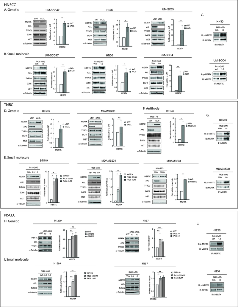
(A, D, H)Genetic inhibition of AXL in HNSCC, TNBC, and NSCLC cell lines. Cells were transfected with AXL siRNA (siAXL) or non-targeting siRNA (siNT). Cell lysates were prepared 72 hours later. (B, E, I) Small molecule inhibition of AXL in HNSCC, TNBC, and NSCLC cell lines. Cells were treated with vehicle or R428 for 72 hours prior to preparation of cell lysates. (F) Antibody inhibition of AXL in TNBC cell lines. Cells were treated with vehicle or Mab173 for 120 hours prior to preparation of cell lysates. Indicated proteins detected by immunoblot. α-Tubulin is shown as a loading control. MERTK expression was quantitated by densitometry using Image J software. Mean values and standard errors derived from 3 independent experiments are shown. *p<0.05, **p<0.01, ns=not significant. (C, G, J) The indicated cell lines were treated with vehicle or R428 for 24 hours and phosphorylated proteins were stabilized by treatment with pervanadate phosphatase inhibitor prior to preparation of cell lysates. MERTK was immunoprecipitated and phosphorylated (denoted by p-) and total MERTK proteins were detected by immunoblot. The data shown are representative of 2 or 3 independent experiments.