
Figure 1. TLR stimulation at the time of T-cell activation in vitro affects expression of PD-1.
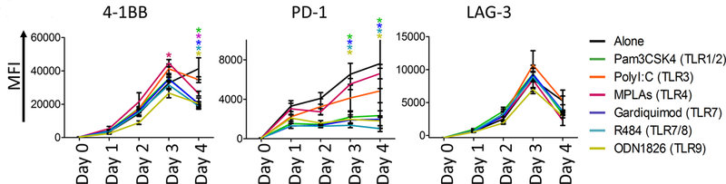
Splenocytes were prepared from the spleens of OT-1 mice and stimulated in vitro with the high-affinity SIINFEKL peptide in the presence or absence of specific TLR agonists [TLR 1/2 (Pam3CSK4), TLR 3 (Poly I:C), TLR 4 (MPLAs), TLR 7 (Gardiquimod), TLR 7/8 (R848), or TLR 9 (ODN1826)]. Shown are the mean fluorescence intensity (MFI) of 4–1BB, PD-1 and LAG-3 on CD8+ T cells collected daily for 4 days. Asterisks indicate p<0.01, with each comparison made to stimulation with SIINFEKL peptide alone, and color corresponding to the agent compared. Results are from one experiment, with samples assessed in triplicate, and are representative of four similar, independent experiments.