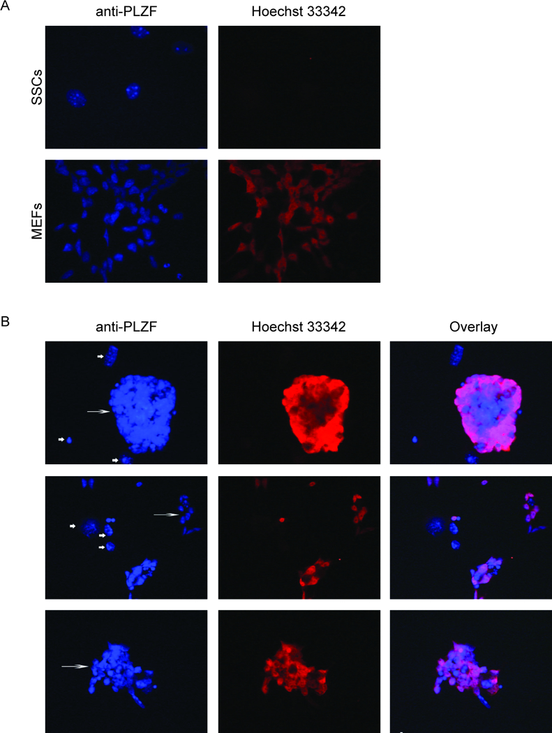
Fig. 2.

Expression of ZBTB16 (PLZF) in MEFs, fresh SSCs, and parental SD-WT2 SSCs kept in culture for 4.5 months (passage 16). Cells were stained with anti-PLZF antibody (red) and Hoechst 33342 (blue). (A) Fresh SSCs (passage 9) on laminin shows undifferentiated spermatogonia that express ZBTB16 (PLZF), whereas the MEFs do not. (B) Immunocytochemistry of SSC clusters at passage 16 on feeders. First column with Hoechst33342 nuclear staining shows SSCs (long arrow) and feeders (short arrow). Second column shows PLZF staining. Third column shows merged images of PLZF staining and Hoechst33342.
