Fig. 5.

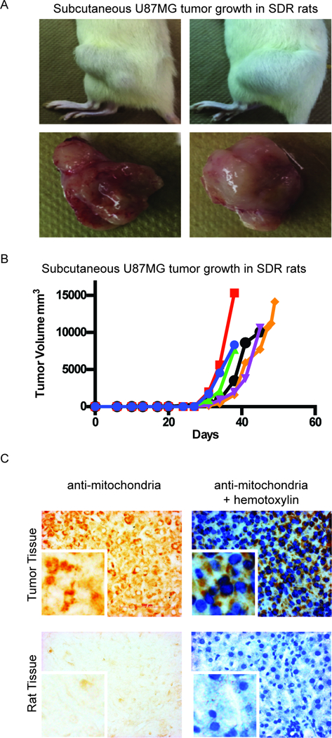
Subcutaneous growth of human glioblastoma U87MG cells in the SDR rat. 1×10⁶ U87MG cells resuspended in Geltrex were injected subcutaneously into SDR rats. (A) Tumor growth in two different SDR animals with images of their excised tumors. (B) Tumor volume (mm3) over time. Each line represents tumor growth in an individual rat. (C) Immunohistochemistry of anti human-mitochondria in tumor tissue and rat tissue. Brown staining demonstrates peri-nuclear localization of human-mitochondria protein in a tumor section, with (right) and without (left) hematoxylin counterstain. 40x magnification. The antibody for human mitochondria protein does not show staining in tissue from a rat that was not injected with human cells (negative control). Right panel with hematoxylin counterstain; 40× magnification; Scale bar = 100μm.
