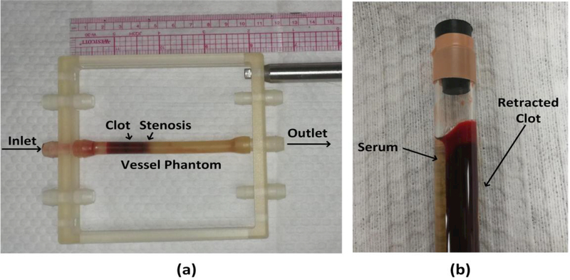
Fig. 2.
(a) The vessel phantom was held by a 3-D-printed frame. A 35% stenosis in the vessel phantom was used to stabilize the clot so that it did not slip under pressure. The inner diameter was 4.2 mm on the downstream side of the stenosis and 6.5 mm on the upstream side. (b) A clot was retracted in a hydrophilic glass tube after 3 d in 4°C incubation.

