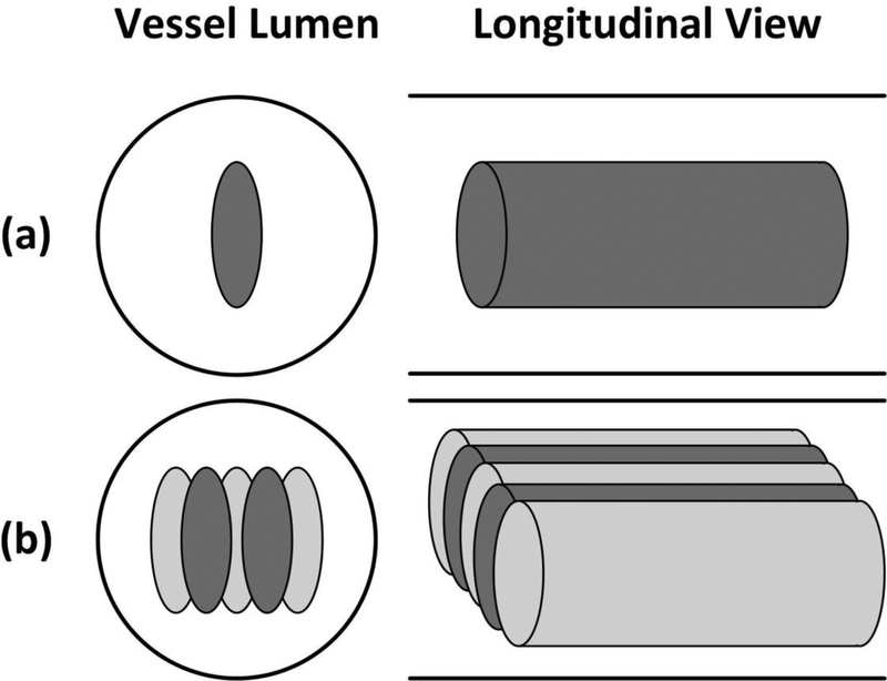
Fig. 5.
Schematic diagrams of the treatment strategies in the vessel phantom, with the solid lines outlined the inner vessel lumen. The ellipses in the vessel lumen represent the treatment foci. (a) The single-focus strategy; (b) the 2+3-foci dual-pass strategy. The darker foci were treated in the first treatment pass and the light grayish foci were treated in the second treatment pass.

