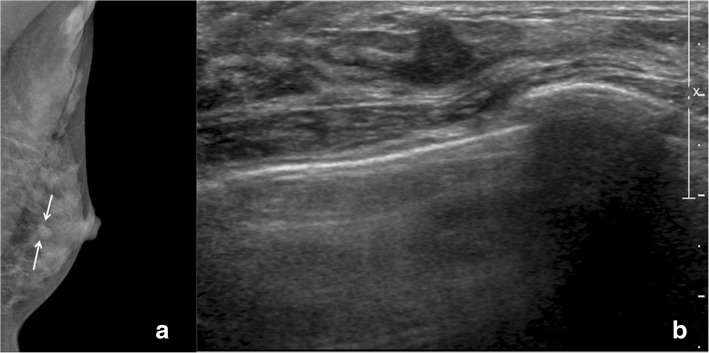
Fig. 2.
A radial scar on ultrasound guided core needle biopsy and upgraded to radial scar with atypical ductal hyperplasia after surgical excision. a On mammography, there is an irregular, indistinct, hyperdense mass (arrow). b On ultrasound, there is an irregular, non-parallel, angular, hypoechoic mass

