Figure 3.

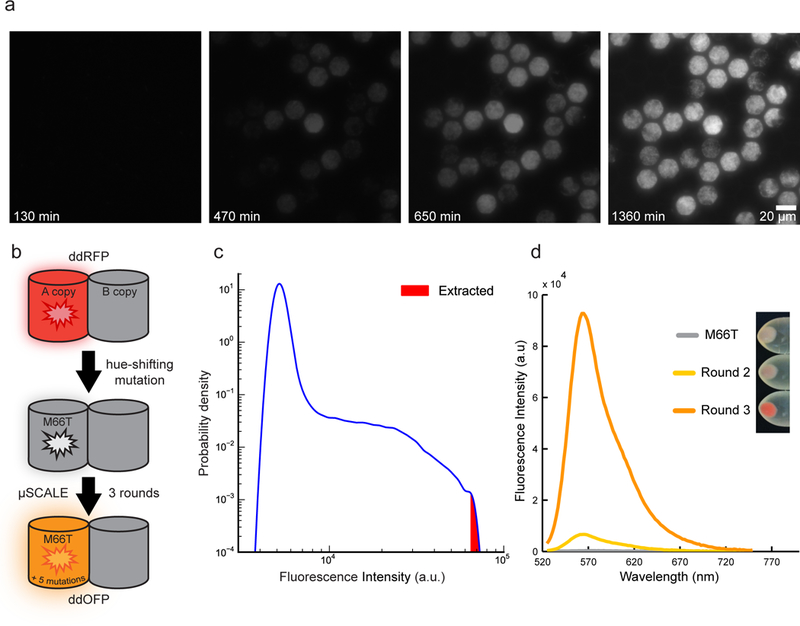
Engineering an orange-hued fluorescent protein using μSCALE. (a) Growth of spatially segregated E. coli cultures expressing GFP. Images depict a small portion of a 20 μm array, which was loaded with, on average, a single bacterial cell per microcapillary, cultured at 37 °C for the indicated times, and serially imaged. (b) Engineering strategy used to generate ddOFP from the ddRFP template. A M66T mutation was introduced into the A copy of ddRFP (starburst), generating a largely non-fluorescent hue-shifted variant. Three rounds of mutagenesis and μSCALE screening of protein libraries expressed in E. coli resulted in ddOFP, which has 5 mutations in addition to M66T. (c) Quantification of μSCALE screen of the third E.coli library. The distribution of observed fluorescence intensities of the library variants is shown in blue (n = 305,279 microcapillaries). The 10 brightest cells at the upper tail of this distribution (red) were extracted from the array. (d) Iterative directed evolution results in variants with increased fluorescence intensity. Plot depicts fluorescence emission spectra of E. coli crude lysates expressing M66T, a variant from round 2, and ddOFP from round 3. Inset depicts the increase in color hue of the cell pellets of E. coli expressing FP variants described above.
