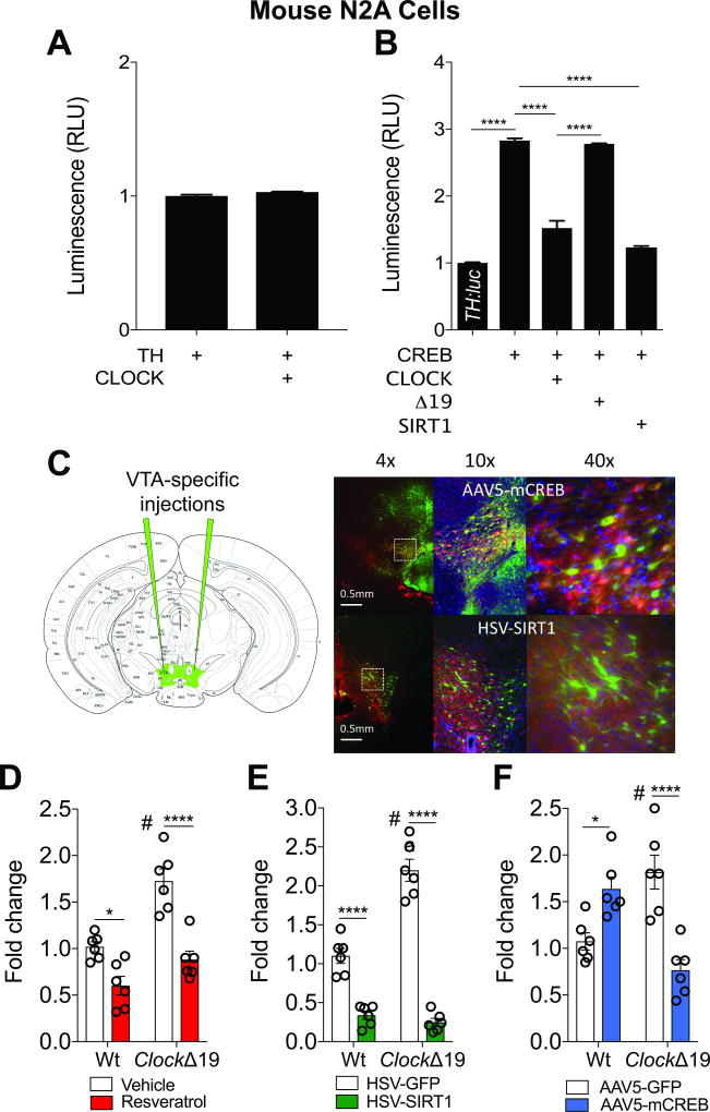
Figure 3. CLOCK and SIRT1 Reduce TH Expression in Neural Cells and In Vivo.
(A) Mouse N2A cells transfected with TH:luc and CLOCK has no effect on bioluminescence activity.
(B) CLOCK and SIRT1 block CREB-induced TH:luc activity in mouse N2A cells (one-way ANOVA, F4,25=279.7, p<0.0001; Tukey’s post hoc tests, **** p<0.0001). n=6 per condition, data represents an average across triplicate wells and multiple plates.
(C) Separate cohorts of mice were injected with HSV-SIRT1 (or GFP, 2–3 days of expression) or AAV5-mCREB (or GFP, 3–4 weeks of expression) into the VTA. Triple-labeling immunohistochemistry shows robust viral expression in the VTA (green, GFP; red, TH; and blue, DAPI).
(D) Resveratrol reduces TH expression in the VTA of Wt and ClockΔ19 mice (genotype, F1,20=25.67, p<0.0001, treatment, F1,20=42.09, p<0.0001, and genotype × treatment, F1,20=4.8, p=0.04; Tukey’s post hoc tests, * p<0.05, **** p<0.0001, # VEH Wt vs. VEH ClockΔ19, p<0.01).
(E) Overexpression of SIRT1 reduces TH expression in the VTA of Wt and ClockΔ19 mice (genotype, F1,20=29.44, p<0.0001, treatment, F1,20=213.4, p<0.0001, and genotype × treatment, F1,20=40.88, p<0.0001; Tukey’s post hoc tests, **** p<0.0001, # VEH Wt vs. VEH ClockΔ19, p<0.0001).
(F) mCREB reduces TH expression only in ClockΔ19 mice (genotype × virus, F1,20=37.05, p<0.0001; Tukey’s post hoc tests, * p<0.05, **** P<0.0001, # AAV5-GFP Wt vs. ClockΔ19, p=0.004).

