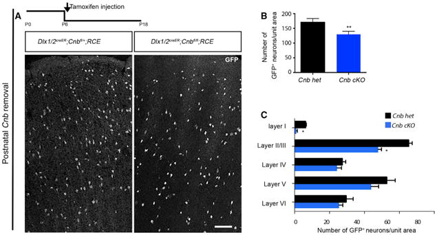
Figure 7. Timing of CaN Removal Is Critical in Regulating Cell Death.

(A) Experimental design and representative image of coronal sections showing the distribution of GFP+ interneurons upon conditional removal of Cnb using an inducible driver line, Dlx1/2creER, reported by EGFP expression by using RCE∷lox P. Tamoxifen was injected at P5, and the brains were analyzed at P18.
(B) Quantification of the number of EGFP+ interneurons shows a reduction in cell density upon conditional removal of Cnb. n = 4, p = 0.0050 (paired t test).
(C) Quantification of the layer distribution using the Dlx1/2creER driver line shows a decrease in the density in superficial layers. Scale bar, 100 μm. See also Figure S7.
