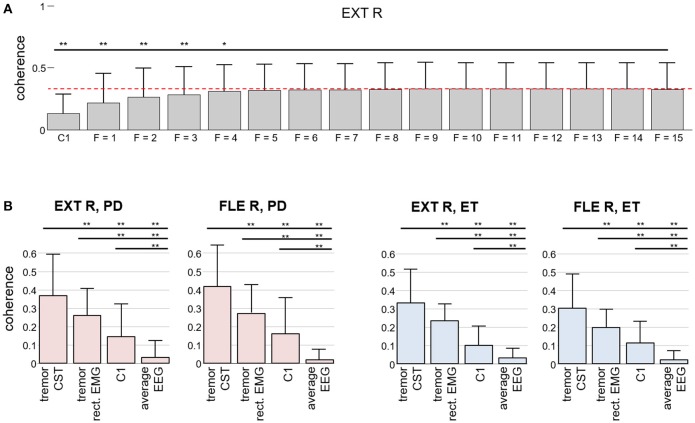
Figure 6.
Comparison of our method for detecting tremor-related cortical activity to the traditional coherence approach in experimental recordings. (A) Coherence between the CST of the right wrist extensor and the estimated tremor EEG component as a function of the extension factor F. For reference, the average coherence with the EEG signals over C1 is also depicted. The results are averaged over all the trials from the PD and ET patients and reported as mean (bars) and SD (whiskers). (B) Coherence between the Laplacian-filtered EEG and the CST in comparison with the coherence between the extracted tremor EEG component and the CST (our proposed method, F = 8): Two results for CST-EEG coherence are depicted in each graph, one for coherence between CST and spatially averaged EEG (“average EEG”) and one for coherence between CST and EEG channel at C1 position (“C1”). Similarly, two results for coherence between CST and extracted tremor component are depicted, one for tremor component, extracted from EEG by using the CST-based estimation described in the Appendix (“tremor CST”) and one for tremor component, extracted from EEG by replacing the CST in Appendix by the spatially averaged rectified EMG, low pass filtered at 15 Hz (“tremor rect. EMG”). Results are plotted separately for each investigated muscle. Superscripts *p < 0.05 and **p < 0.01 denote statistically significant difference as assessed by Wilcoxon signed rank test.

