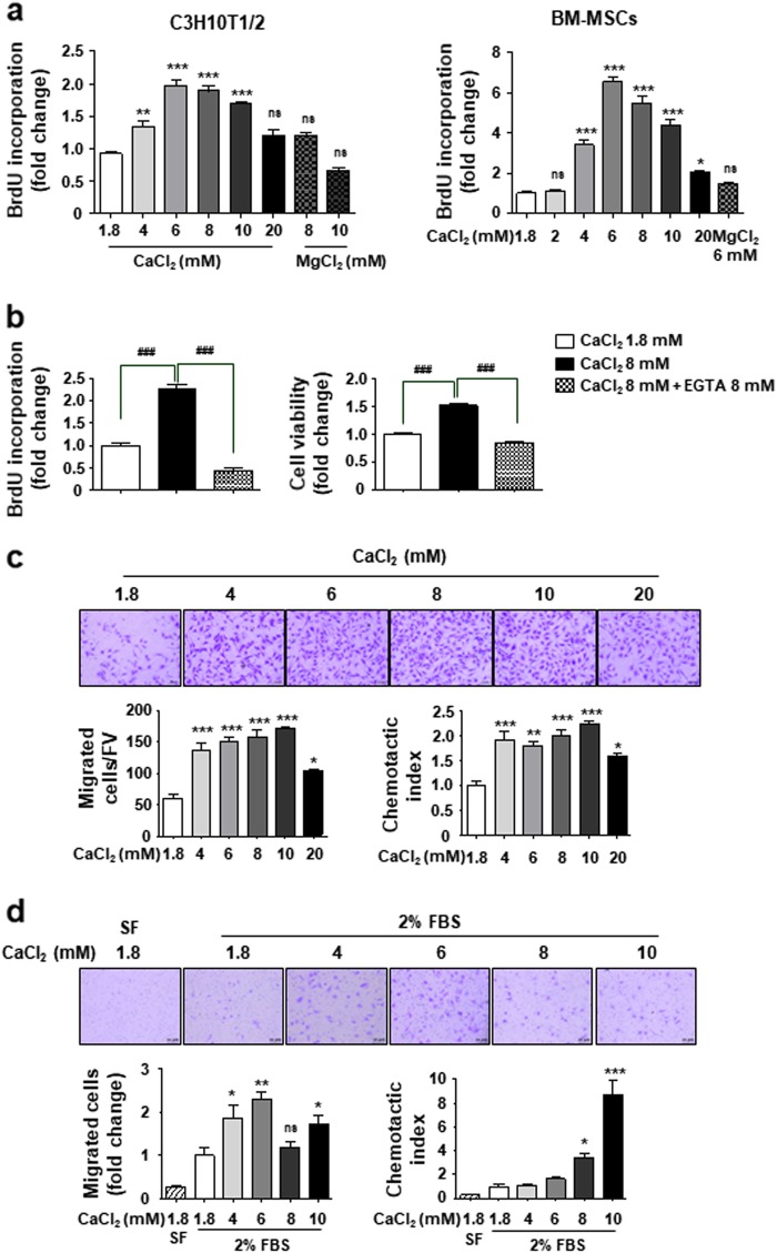
Fig. 1. Effect of extracellular Ca2+ on MSCs behavior.
a, b Extracellular Ca2+ promotes cell proliferation in a concentration-dependent manner. Cells were cultured in 2% FBS-containing medium for 48 h with the indicated amounts of either CaCl2 or MgCl2. EGTA was used as a Ca2+ chelator. Cell proliferation was measured by the BrdU incorporation assay and cell viability was determined by CCK-8 assay. c, d Extracellular Ca2+ promotes cell migration in a concentration-dependent manner. C3H10T1/2 cells (c) and BM-MSCs (d) were used in Transwell assay with the indicated amounts of CaCl2-containing medium in the lower chamber. Cells that migrated to the lower side of the well for 24 h were fixed and stained. The number of migrated cells per field of view (FV, x50 magnification) is indicated (bottom left in c). The number of migrated cells was expressed as the fold increases relative to control (bottom left in d). The migration capacity of cells was expressed as the chemotatic index by correcting the effect of Ca2+ on the proliferation of migrated cells during the 24 h treatment (chemotatic index = migrating cell number/fold change of proliferating cells, bottom right). SF (serum-free medium), negative control. *p < 0.05; **p < 0.01; ***p < 0.001 vs. first bar, #p < 0.05; ##p < 0.01; ###p < 0.001 vs. indicated group

