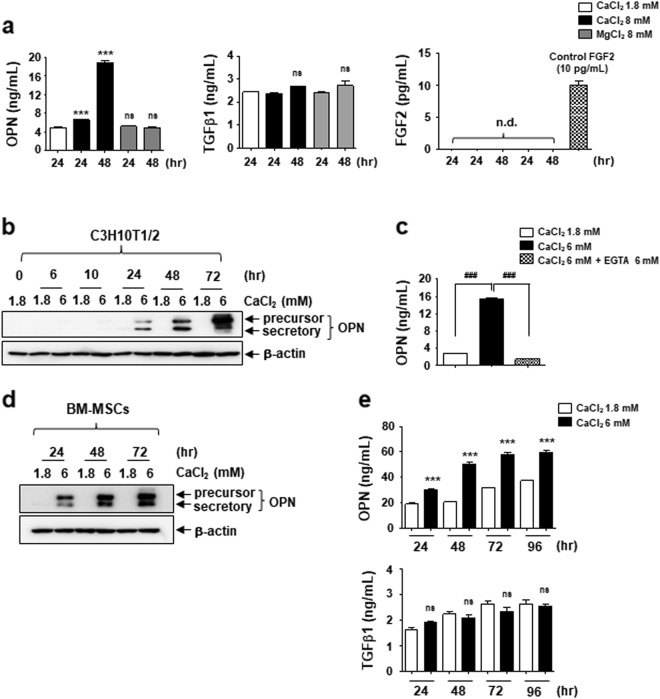
Fig. 3. Extracellular OPN is increased by MSCs under elevated extracellular Ca2+ conditions.
a Measurement of extracellular OPN, TGFβ1, and FGF2 in the indicated conditioned medium. Conditioned medium was harvested from different cultures of C3H10T1/2 cells as indicated and used to ELISA as described in the Materials and methods section. b and d Time course of effect of elevated extracellular Ca2+ on OPN protein levels. C3H10T1/2 cells (b) and BM-MSCs (d) were treated with 6 mM Ca2+ medium and harvested at the indicated time points followed by Western blotting analysis. c The expression and secretion of OPN is induced by elevated extracellular Ca2+ specifically. Extracellular OPN levels were analyzed by ELISA after the C3H10T1/2 cells were incubated for 72 h as indicated. e Extracellular OPN and TGFβ1 level in the conditioned medium derived from different cultures of BM-MSCs as indicated. ***p < 0.001 vs. first bar. ###p < 0.001 vs. indicated group. ns not significant; nd not detected

