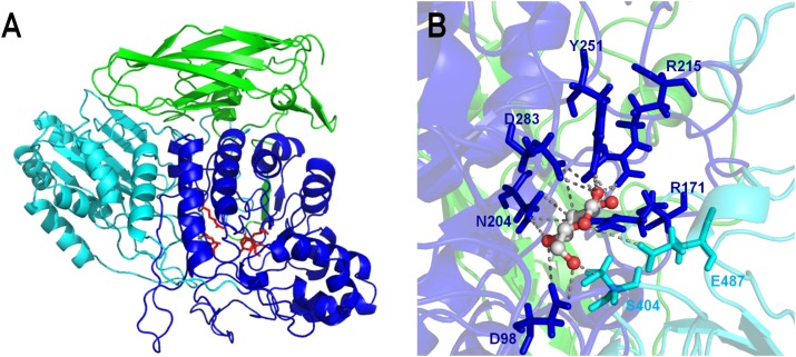
FIGURE 3.
3D-structure model of the β-glucosidase Lfa2 characterized in this work. (A) Overall 3D-structure representation showing the three domains of the β-glucosidase: TIM barrel-like domain (in blue), α/β sandwich domain (in cyan) and the C-terminal fibronectin type III domain (in green). Side chains of the catalytic amino acids and amino acids involved in substrate binding are shown in red. (B) Catalytic site representation in complex with glucose. Amino acid side chains are represented by sticks, indicating the conserved catalytic amino acids D283 and E487 and other amino acids involved in substrate recognition and stabilization (D98, R171, N204, R215, Y251, and S404).

