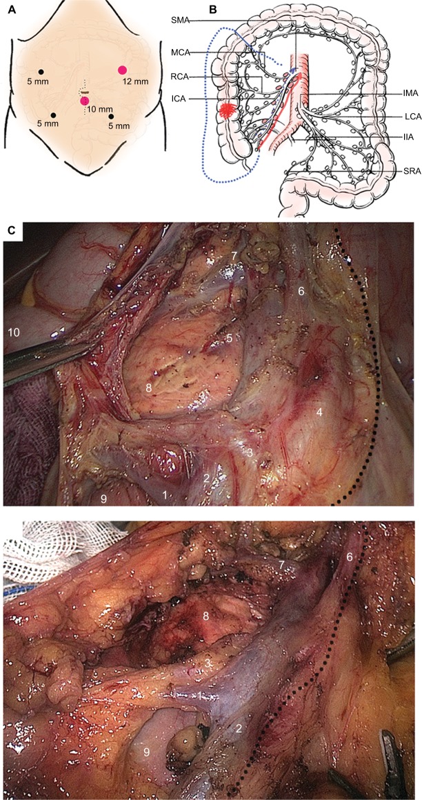
Figure 1.
Demonstration of D3 lymphadenectomy for laparoscopic right hemicolectomy.
Notes: (A) Position of five operative ports; (B) Scope of D3 lymphadenectomy according to various surgical methods. The direction following the red arrow indicates the SLRC method, whereas that direction following the blue dotted arrow indicates the conventional SMV-guided LRC method. The blue short line indicates the cutting edge of distal bowel. (C) The typical D3 lymphadenectomy and vessel skeleton on basis of SLRC technique. LN dissection was started from the inside of SMA with a caudal-to-cranial medial approach (the dotted line). Several tributaries of SMV and SMA were ligated after sufficient space dissection (1. Ileocolic vein; 2. SMV trunk or surgical trunk; 3. Root of ileocolic artery; 4. SMA trunk; 5. Superficial dorsal vein of pancreas; 6. Root of arteria colica media; 7. Henle trunk, also known as gastrocolic trunk; 8. Head of pancreas; 9. Horizontal part of duodenum; 10. Hepatic flexure). (D) The typical D3 lymphadenectomy and vessel skeleton on basis of CLRC technique. The SMA is not exposed routinely by using this technique. The dotted line stands for the inside region of lymph node dissection beyond the SMV. Those numbers indicate vessels and organs (1. Ileocolic vein; 2. SMV trunk or surgical trunk; 3. Root of ileocolic artery; 6. Root of arteria colica media; 7. Henle trunk, also known as gastrocolic trunk; 8. Head of pancreas; 9. Horizontal part of duodenum).
Abbreviations: CLRC, conventional laparoscopic right hemicolectomy; ICA, ileocolic artery; IIA, inner iliac artery; IMA, inferior mesenteric artery; LCA, left colic artery; LRC, laparoscopic right hemicolectomy; MCA, middle colic artery; RCA, right colic artery; SLRC, SMA-guided LRC; SMA, superior mesenteric artery; SMV, superior mesenteric vein; SRA, superior rectal artery.

