Figure 1.
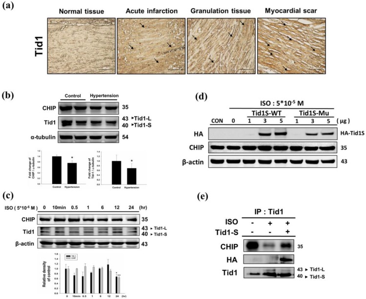
Expression Tid1 and CHIP in different stage of myocardial infarction tissues and H9c2 cardiomyoblast cells treated with ISO. (a) Immunohistochemical analysis, with the indicated antibodies, of serial sections of representative lesions. Tid1 accumulations were increased with disease progression visualized dark-brown color which indicated with arrows. Panels: a normal tissue (NT, n=10), an acute infraction (AI, n=10), a granulation tissue (GT, n=10) and a myocardial scar (MS, n=10). Final magnifications: × 200 (bar, 200 μm). (b) The expression of CHIP and Tid1 in hypertension models were measured via immunoblotting. Quantification of the results is shown blow from three independent experiments; mean ± S.D., * P < 0.05. (c) H9c2 cells were treated with different dosages of ISO (5*10-5 M) in serum-free medium for 24 hours were measured via immunoblotting. Quantification of the results is shown blow from three independent experiments; mean ± S.D., * P < 0.05, ** P < 0.01. (d) H9c2 cells were pretreated with ISO (5*10-5 M) in serum-free medium for 24 hours, and then transfected with different amounts of Tid1S-WT (1, 3 and 5μg) and Tid1Su plasmid (1, 3 and 5μg) for 24 hours. Cell was harvest and analyzed via immunoblotting. (e) H9c2 cells were pretreated with ISO (5*10-5 M) then transfected with Tid1S-WT plasmid and harvested for immunoprecipitation with anti Tid1 antibody, and the samples were analyzed via immunoblotting.

