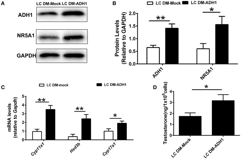
Figure 4.
Overexpression of ADH1 promotes mESC differentiation into Leydig cells. (A,B) Representative Western blot for protein expression of ADH1 and NR5A1 in SF1-overexpressing mESCs (mESCs-SF1) transfected with adenovirus (empty vector or Adh1) at day 6. Relative protein expression levels were normalized to GAPDH. (C) Analysis of the expression of key genes involved in steroidogenic enzymes in mESCs-SF1 transfected with empty vector or Adh1. (D) Testosterone production in different culture conditions at day 6. All quantitative data were obtained from three independent experiments and are presented as mean ± SD; **P < 0.01 and *P < 0.05. LC DM-Mock, mESCs-SF1 transfected with empty vector, then cultured in Leydig cell differentiation medium (LC DM) for 6 days. LC DM-ADH1, mESCs-SF1 transfected with Adh1, then cultured in LC DM for 6 days.

