Figure 2.

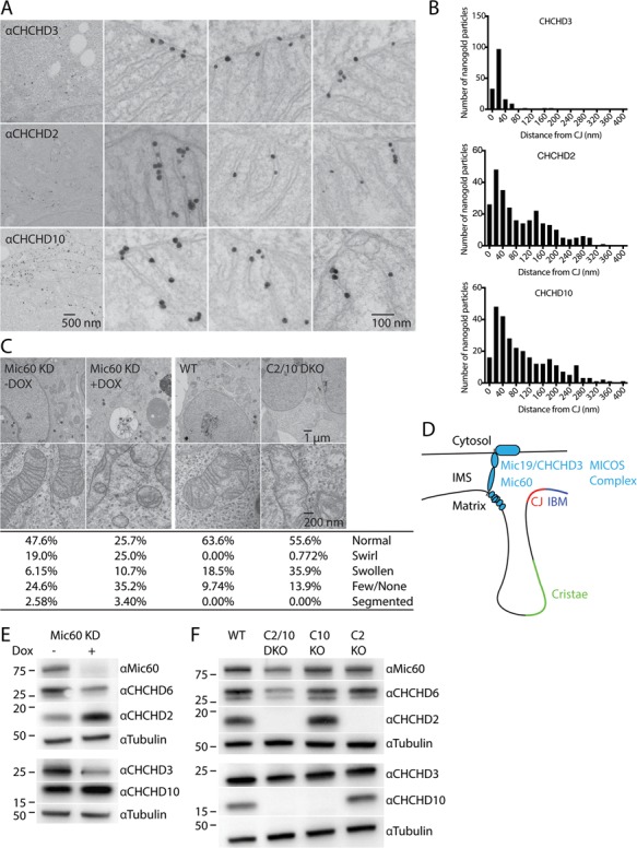
CHCHD2 and CHCHD10 localize to mitochondria cristae and are not essential for MICOS complex stability. (A) Representative Immuno-TEM images labelled pre-embedding with MIC19/CHCHD3, CHCHD2 or CHCHD10 antibodies followed by nanogold-conjugated secondary antibodies. (B) Histograms depicting distance from individual intra-mitochondrial nanogold particles to the nearest cristae junction (CJ) for MIC19/CHCHD3 (top), CHCHD2 (middle) and CHCHD10 (bottom). Particles in the cytosol or associated with the cytosolic face of the outer membrane were excluded from the analysis. (C) Representative TEM images from Doxycycline (Dox) inducible knockdown HeLa cells targeting MIC60 (MIC60 KD), WT HEK293 cells and C2/10 DKO cell lines. Dox-inducible cell lines were treated with 2 μm Dox for 7 days (Dox+) or left untreated (Dox−). Greater than 200 mitochondria were scored per condition. (D) Model of MICOS complex relative to the intermembrane space (IMS) and inner membrane composed of the inner boundary membrane (IBM), CJ and cristae. (E) Lysates from MIC60 KD HeLa cells +/− Dox were immunoblotted with MIC60, MIC25/CHCHD6, CHCHD2, MIC19/CHCHD3, CHCHD10 and Tubulin antibodies. Tubulin served as a loading control. (F) Lysates from WT, C2 KO, C10 KO, C2/10 DKO HEK293 cells were immunoblotted with the same antibodies as in (E).
