
Figure 3.
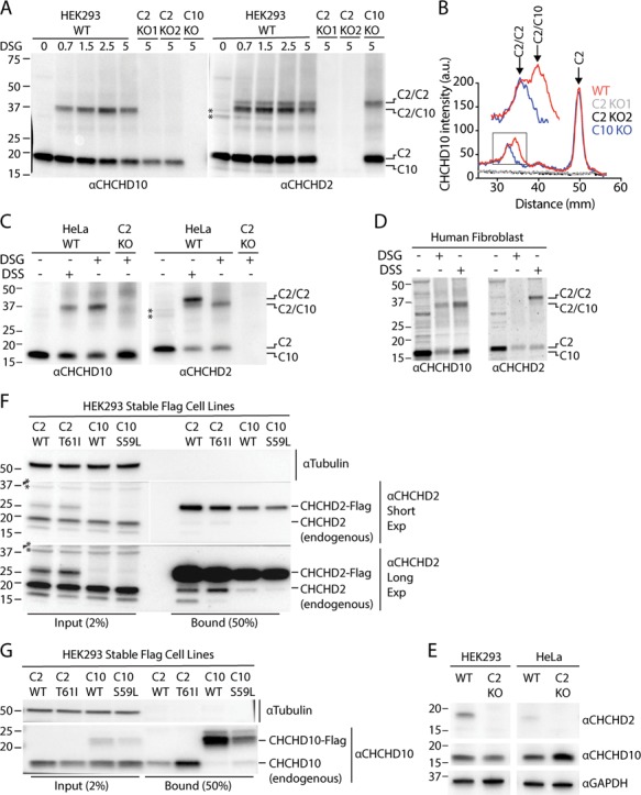
CHCHD2 promotes oligomerization of endogenous CHCHD10. (A) Lysates from WT, C2 KO and C10 KO HEK293 cells treated with the indicated DSG concentration or untreated were immunoblotted with CHCHD2 and CHCHD10 antibodies. Two independent C2 KO cell lines (C2 KO1 and C2 KO2) were tested. (B) Representative line scans of experiment described in (A). Experiment was performed >= 3 times on at least two separate occasions with similar results. (C) Lysates from untreated, DSG (5 mm) or DSS (5 mm) treated WT and C2 KO HeLa cells were immunoblotted with CHCHD2 and CHCHD10 antibodies. (D) Lysates from untreated, DSG (5 mm) or DSS (5 mm) treated primary human fibroblasts were immunoblotted with CHCHD2 and CHCHD10 antibodies. (E) Lysates from HEK293 and HeLa WT and C2 KO cells were immunoblot ted with antibodies against CHCHD2, CHCHD10 and GAPDH. (F and G) Lysates from HEK293 cells stably expressing CHCHD2 WT-Flag (C2 WT), CHCHD2 T61I-Flag (C2 T61I), CHCHD10 WT-Flag (C10 WT) and CHCHD10 S59L-Flag (C10 S59L) were immunocaptured with anti-Flag beads, followed by immunoblotting with Tubulin, CHCHD2 (F) and CHCHD10 (G) antibodies. Tubulin was used as a loading control. Experiment was performed >= 3 times on at least two separate occasions with similar results. In all panels ‘*’ indicates two non-specific bands cross-reactive with the mouse monoclonal CHCHD2 antibody that was used in all panels of the figure. These were observed in untreated but not in DSG or DSS treated samples.