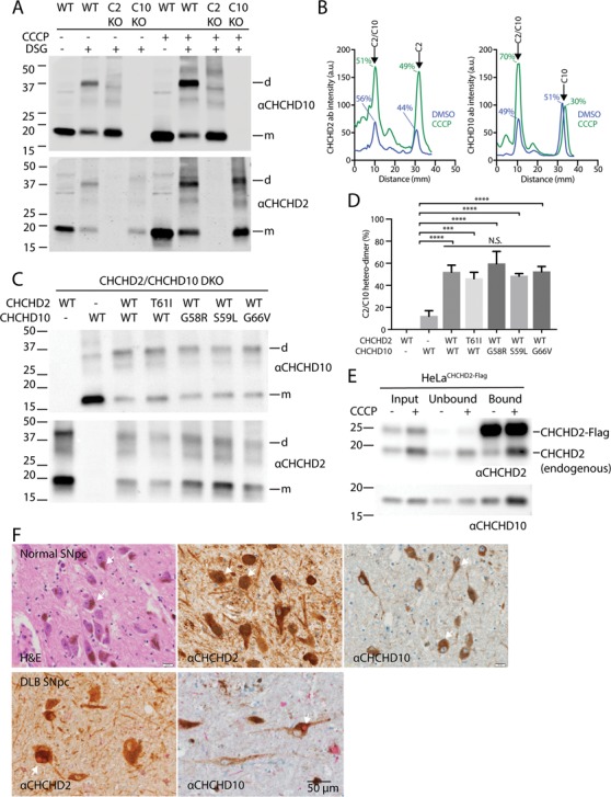
Figure 6.

Loss of Δψ promotes CHCHD2/10 heterodimerization, and CHCHD2/10 with disease-causing mutations readily incorporate into heterodimers. (A) Lysates from HeLa cells treated with DMSO or CCCP (10μM) for 24 h immunoblotted for CHCHD2 and CHCHD10. (B) Line scan of immunoblot in (A). (C) Lysates from CHCHD2/10 DKO HEK293 cells co-transfected with the indicated CHCHD2 and CHCHD10 WT or missense mutation-containing cDNAs were treated with DSG (5mM). In control experiments (lanes 1–2), CHCHD2 or CHCHD10 was co-transfected with YFP cDNA. (D) Quantification of experiment described in (C). Intensity of CHCHD10 antibody signal was determined by line-scanning in three biological replicates. The percent dimer represented the ratio of the peak corresponding to the dimer divided by the sum of the dimer and monomer peaks. One-way analysis of variance (ANOVA) followed by Tukey’s multiple comparisons test was used to determine statistical significance. ‘n.s.’, not significant; ‘****’, P-value < 0.0001; and ‘***, P-value = 0.0001. (E) Lysates from HeLa cells stably expressing CHCHD2 WT-Flag (C2 WT) and treated with CCCP or DMSO overnight were immunocaptured with anti-Flag beads, followed by immunoblotting (IB) with CHCHD2 (top) and CHCHD10 (bottom) antibodies. Experiment was performed >= 3 times on at least two separate occasions with similar results. (F) Representative images from normal human substantia nigra pars compacta (SNpc) stained with hematoxylin and eosin (left panel), CHCHD2 antibody (middle panel) and CHCHD10 antibody (right panel). CHCHD2 and CHCHD10 stain are lighter brown than the neuromelanin pigment (arrows) characteristic of dopaminergic neurons in the adult human SNpc. Representative images of SNpc from a DLB case co-immunostained for CHCHD2 (brown) and α-synuclein (red) (left panel) or CHCHD10 (brown) and α-synuclein (red) (right panel). Arrows indicate characteristic brainstem Lewy bodies.
