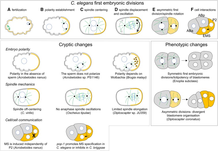
Figure 2.
First embryonic cell divisions in C. elegans and variations in other species. Top panel (A–F) schematic representation of the two first cell division of the C. elegans embryo. Microtubules are shown in green, centrosomes are represented by black dots and nuclei by white circles. Polarity proteins are shown in gray and yellow. (A) Initially, the oocyte is unpolarized. After the sperm entry (on the right), female meiosis resumes (spindle on the left). (B) After fertilization, polarity proteins are asymmetrically localized and the sperm entry site defines the posterior pole of the cell on the right (B). In response to polarity, the mitotic spindle (D) and cell fate determinants (E) are asymmetrically localized. During the second cell division, spindle orientation is different between the two cells (E), giving rise to a rhomboid organization of blastomeres at the four-cell stage (F). At this stage, the P2 cell sends a Wnt signal to EMS. Phenotypic changes: timing of cell divisions and cell orientations can vary between species leading to different cellular contacts and blastomeres organization. Cryptic changes: among species that have similar embryonic cell divisions than C. elegans, evolutionary changes are found in the polarization of the embryo, the positioning of the first mitotic spindle or in cell/cell communication.

