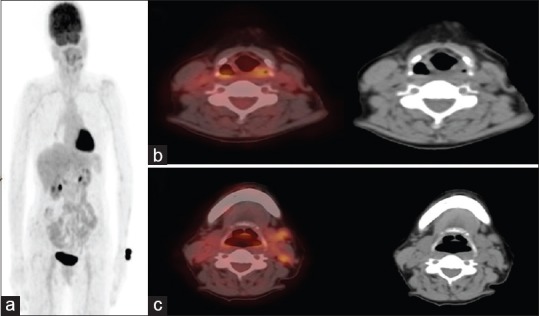
Figure 1.

18F-fluorodeoxyglucose-positron emission tomography/computed tomography in 52 year old female with fine-needle aspiration cytology proven metastatic cervical lymph nodes. (a) Whole body maximum intensity projection image. (b) Axial fused positron emission tomography/computed tomography and computed tomography image showing luorodeoxyglucose avid thickened left aryepiglottic fold, subsequent guided biopsy confirmed it as primary site. (c) Axial fused positron emission tomography/computed tomography and computed tomography image showing fluorodeoxyglucose avid metastatic left cervical lymph nodes
