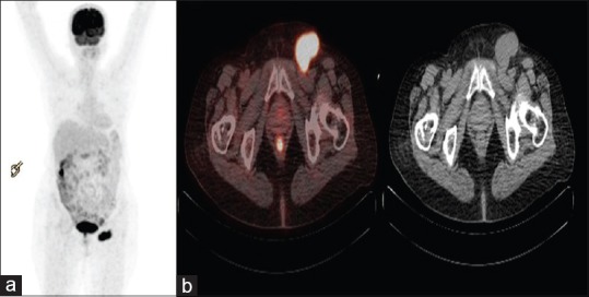
Figure 2.

18F-fluorodeoxyglucose-positron emission tomography/computed tomography in 55 year old female with biopsy proven metastatic left inguinal lymph nodes. (a) Whole body positron emission tomography maximum intensity projection image showing abnormal fluorodeoxyglucose uptake in left inguinal region. (b) Axial Fused positron emission tomography/computed tomography and computed tomography image in metastatic left inguinal lymph nodes. Primary lesion could not be detected
