Figure 2.
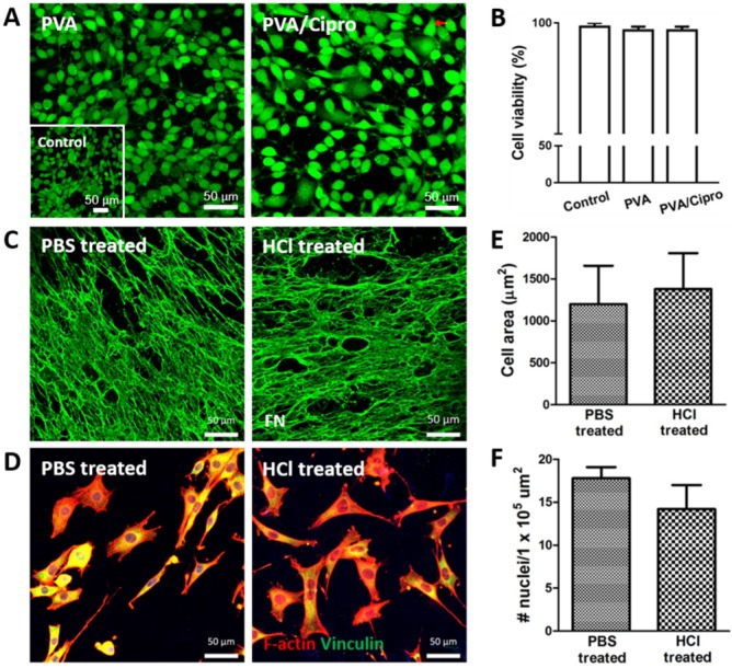
Characterization of PVA-based membranes. (A) Cell viability test of mouse fibroblasts after 24 h culture on gelatin coated-PVA (treated with 0.1 N HCl) and gelatin-coated PVA/Cipro (treated with 1 mg/mL ciprofloxacin) as evaluated via live/dead assay. Inset shows the cells cultured on TCP. (B) Quantitative analysis of cell viability based on image analysis of the live/dead assay. (C) Immunofluorescence of hFDM against FN in PVA/hFDM treated with either PBS or 0.1 N HCl. (D) Culture of fibroblasts on PBS-treated and 0.1 N HCl-treated PVA/hFDM, respectively. F-actin is stained red and focal adhesion molecule vinculin is shown in green. (E) Quantitative analysis of cell spreading area and (F) cell density based on image analysis of (D). All scale bars are 50 μm. Cipro: ciprofloxacin; FN: fibronectin; HCl: hydrochloric acid; hFDM: human lung fibroblast-derived matrix; PBS: phosphate buffered saline; PVA: polyvinyl alcohol; TCP: tissue culture plastic.

