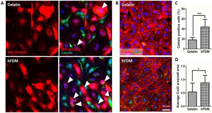
Figure 4.
Gap junction-mediated cell-cell communication. (A) Dye transfer assay between calcein AM-labeled HUVECs (green) and RFP-HUVECs (red) after 24 h co-culture on gelatin and hFDM, respectively; the images in the left side show only the RFP-HUVECs, and the merged images of RFP-HUVECs and calcein AM-labeled HUVECs are on the right side. Positive signals (white triangles) are identified via the RFP-HUVECs (red) with green dye (calcein AM) uptake. (B) Expression of Cx43 protein (green) with HUVECs cultured on either gelatin or hFDM for 3 days, along with F-actin staining (red). (C) Percentage of calcein dye-positive RFP-HUVECs as calculated via image analysis after the dye transfer assay; the percentage was averaged by counting the number of dye-positive RFP-HUVECs against total RFP-HUVECs in a given image. (D) Quantitative evaluation of average Cx43 area per cell area. All scale bars are 50 μm. Statistically significant difference: *p < 0.05 or ***p < 0.001. Cx43: connexin 43; HUVECs: human umbilical vein endothelial cells; RFP-HUVECs: red fluorescence protein expressing HUVECs.

