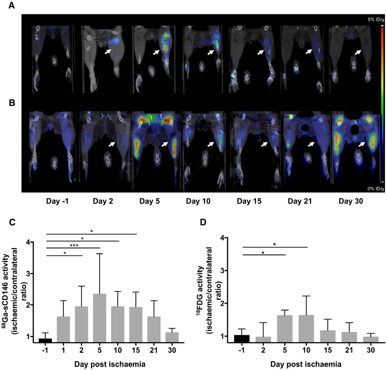
Figure 5.
68Ga-sCD146 and 18F-FDG imaging after hindlimb ischaemia. (A) Representative 68Ga-sCD146 PET images of a hindlimb ischaemia model. (B) Representative 18F-FDG PET images of a hindlimb ischaemia model (white arrow: ischaemic hindlimb). (C) 68Ga-sCD146 hindlimb uptake 40 min post-injection expressed as ischaemic-to-contralateral muscle ratio on days 1, 2, 5, 10, 15, 21 and 30 post-ischaemia (*P < 0.05 vs D-1; ***P = 0.0005 vs D-1; n = 7). (D) Hindlimb uptake 40 min post-injection of 18F-FDG expressed as ischaemic-to-contralateral muscle ratio on days -1, 2, 5, 10, 15, 21 and 30 post-ischaemia (*P < 0.05 vs D-1; n = 5).

