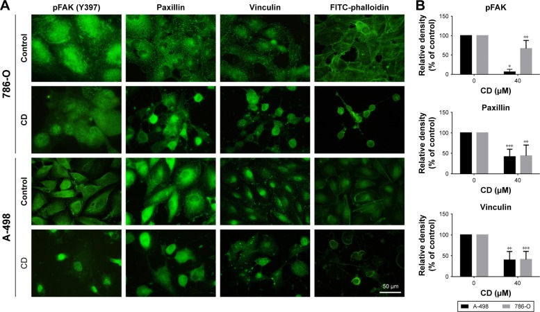
Figure 5.
RCC cells were treated with 0.1% DMSO (control) or 40 µM CD for 24 hours.
Notes: (A) Immunofluorescence staining was used to detect FA proteins. Stress fibers were determined by staining with FITC-phalloidin. (B) The bar graphs show densitometric data from four to eight different coverslips. Scale bar =50 µm. *P<0.05, **P<0.01, and ***P<0.001.
Abbreviations: CD, 16-hydroxycleroda-3,13-dien-15,16-olide; DMSO, dimethyl sulfoxide; FA, focal adhesion; FITC, fluorescein isothiocyanate; pFAK, phosphorylated focal adhesion kinase; RCC, renal cell carcinoma.

