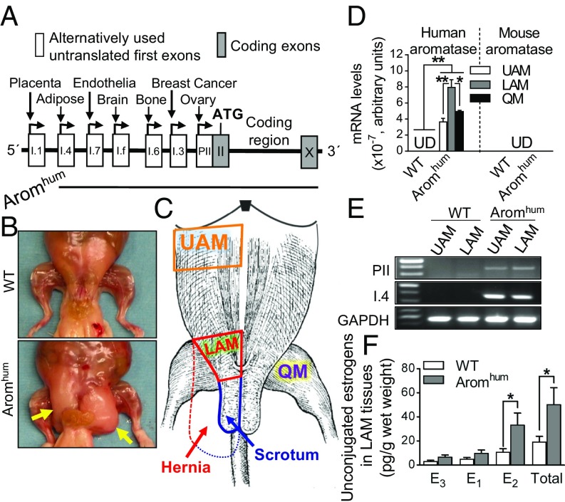
Fig. 1.
Human aromatase expression, abdominal muscle tissue estrogen levels, and scrotal hernia formation in Aromhum transgenic mice. (A) Schematic of the human BAC clone construct used to generate Aromhum transgenic mice, which contained all alternatively used promoters with downstream first exons of the aromatase gene except for placental exon I.1. (B) Dissected abdominal musculature of 26-wk-old WT and Aromhum mice. Yellow arrows indicate scrotal hernia. (C) Schematic of mouse abdominal muscle anatomy in WT and Aromhum mice. UAM, LAM, QM, the scrotum, and hernia are shown in the sketch. Solid red line and solid blue line indicate normal LAM tissue and normal scrotum in WT mice, respectively. Dashed red line indicates expanded fibrotic LAM tissue that comprises the hernia wall contiguous with the scrotum (dashed blue line) in Aromhum mice. (D) Human and mouse aromatase mRNA levels were determined in UAM, LAM, and QM of Aromhum mice. UD, undetermined. Statistical analysis by two-way ANOVA with Sidak’s multiple comparison test, *P < 0.05, **P < 0.01, n = 8 mice per group. (E) Exon-specific RT-PCR confirmed that distinct promoters drive human aromatase expression in the abdominal muscle tissues of Aromhum mice. GAPDH mRNA levels served as internal control. Data are representative of three independent experiments. (F) LAM tissue unconjugated estrogens were measured by LC-MS2 assay. E1, E2, and E3 are shown. Two-tailed Student’s t test, *P < 0.05, n = 14 mice.

