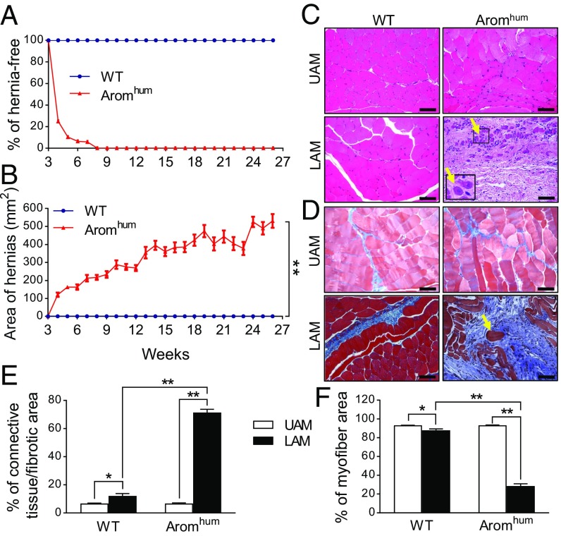
Fig. 2.
Human aromatase expression and estrogen production lead to LAM tissue fibrosis, myocyte atrophy, and the development of scrotal hernia in Aromhum mice. (A) The incidence of scrotal hernia. Scrotal hernia formation was monitored by weekly visual inspection from 3 to 26 wk of age. No scrotal hernia was seen in the WT group. n = 32 mice. (B) The area of scrotal hernia was measured from 3- to 26-wk-old mice. Two-tailed Student’s t test, **P < 0.01 for Aromhum vs. WT, n = 32 mice. (C) H&E and (D) Masson’s trichrome staining of UAM and LAM from 26-wk-old WT and Aromhum mice. n = 10 mice for C and D. Yellow arrows in C and D indicate one of many atrophied myocytes. (Scale bars, 100 μm; Magnification: Inset in C, 40×.) Quantification of the percentage of connective tissue or fibrotic area (E) or myofiber area (F) in WT and Aromhum mice. Ten representative high-power fields were analyzed in each tissue. Two-way ANOVA with Tukey’s multiple comparison test, *P < 0.05, **P < 0.01, n = 8 mice per group.

