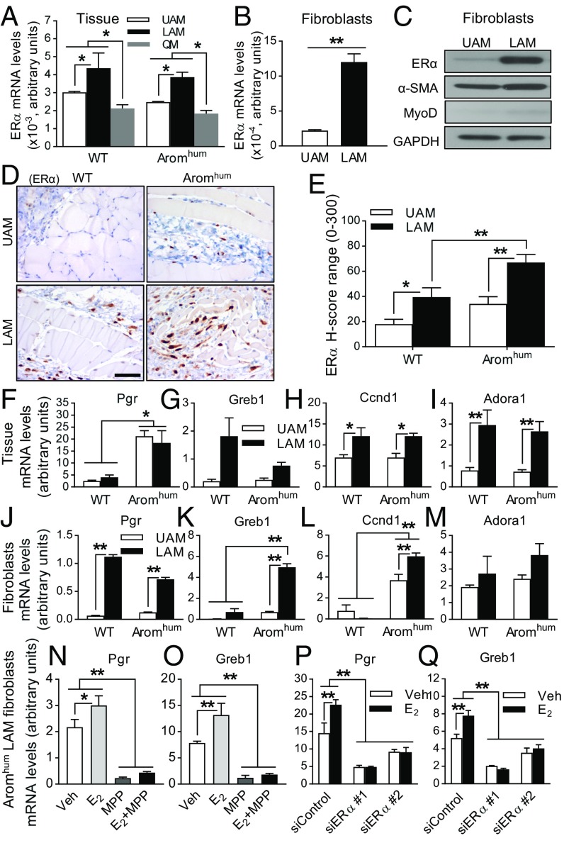
Fig. 4.
ERα expression is higher in LAM tissue fibroblasts of both WT and Aromhum, which contributes to the higher estrogen-responsive gene expression in LAM tissue of Aromhum mice. (A) Relative mRNA levels of ERα in UAM, LAM, and QM of WT and Aromhum mice. Two-way ANOVA with Tukey’s multiple comparison test. *P < 0.05. n = 10 mice in each group. (B) ERα mRNA levels and (C) ERα protein levels in primary fibroblasts from UAM and LAM of WT mice. The data shown in C are representative of three independent experiments. Two-tailed Student’s t test, **P < 0.01, n = 3. (D) ERα localization in UAM and LAM of WT and Aromhum mice was measured by IHC staining. (Scale bar, 50 μm.) (E) A minimum of 1,000 nuclei were counted in sections to calculate the average H-score of ERα in fibroblasts of UAM and LAM from WT and Aromhum mice. Two-way ANOVA with Sidek’s multiple comparison test, *P < 0.05, **P < 0.01, n = 10. mRNA levels of Pgr, Greb1, Ccnd1, and Adora1 in tissue lysates (F, G, H, and I; n = 9) and primary fibroblasts (J, K, L, and M; n = 6) of UAM and LAM from WT and Aromhum mice. mRNA levels of Pgr and Greb1 in LAM primary fibroblasts from Aromhum mice after MPP (10 µM) treatment (N and O) or siRNA-mediated knockdown of ERα (P and Q) in the presence or absence of E2 (10 nM). Cells were pretreated with MPP for 2 h before the addition of E2. Veh, vehicle. Two-way ANOVA with Sidak’s multiple comparison test, *P < 0.05, **P < 0.01. GAPDH mRNA or protein levels served as controls.

