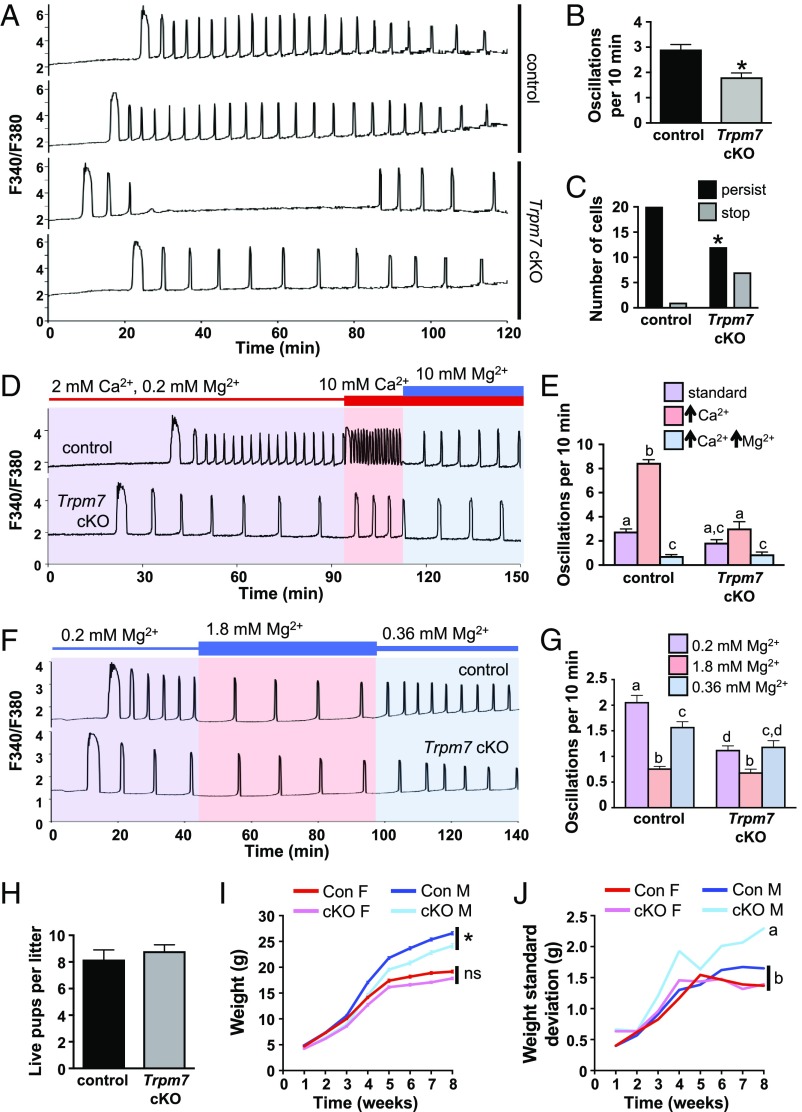
Fig. 3.
Loss of TRPM7 reduces Ca2+ oscillation frequency and persistence in mouse eggs and causes abnormal growth trajectory in male offspring. (A–C) Ratiometric Ca2+ imaging of n = 22 control and 19 Trpm7 cKO eggs was performed during IVF in three independent experiments. (A) Representative traces. (B) Oscillation frequency, mean ± SEM, *P < 0.05, t test. (C) Number of eggs with persistence of Ca2+ oscillations after 60 min. *P < 0.05, Fisher’s exact test. (D and E) Response of n = 18 control and 13 Trpm7 cKO eggs to the indicated changes in Ca2+ and Mg2+ concentration in three independent experiments. (D) Representative traces. (E) Graph of oscillation frequency during incubation in the indicated conditions, mean ± SEM. Different letters above columns indicate significant difference, P < 0.05, two-way ANOVA with Tukey’s multiple comparisons test. (F and G) Response of n = 29 control and 33 Trpm7 cKO eggs to the indicated changes in Mg2+ concentration in three independent experiments. (F) Representative traces. (G) Graph of oscillation frequency during incubation in the indicated conditions, mean ± SEM. Different letters above columns indicate significant difference, P < 0.05, two-way ANOVA with Tukey’s multiple comparisons test. (H–J) Breeding trial of wild-type males bred for 6 mo to females carrying either control or Trpm7 cKO oocytes; seven breeding pairs per group. (H) Average number of live pups per litter, mean ± SEM. (I) Weight of all male and female offspring over time, mean ± SEM, *P < 0.05, mixed-model ANOVA. ns, not significant. N = 24 female pups, 28 male pups from control dams; 32 female pups, 30 male pups from cKO dams. (J) SD of offspring weight over time. Different letters indicate statistically different SDs between groups (mixed-model ANOVA).

