Fig. 5.
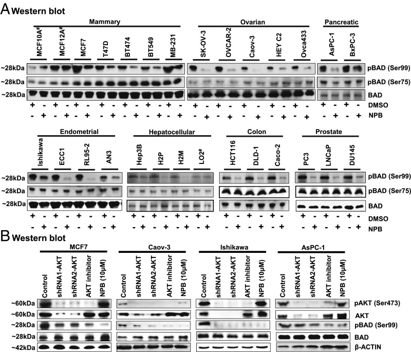
NPB specifically inhibits BAD phosphorylation at Ser99 in carcinoma cell lines independently of AKT signaling. (A) WB analysis was used to assess the levels of phosphorylated hBAD at Ser75 and Ser99 and BAD protein in a range of carcinoma cell lines, including mammary, ovarian, pancreatic, endometrial, hepatocellular, colon, and prostate cancer, after treatment with NPB (5 µM). Total BAD was used as an input control for cell lysate. (B) WB analysis was used to assess the levels of pBAD at Ser99, pAKT at Ser473, AKT, and BAD in MCF7, Caov-3, Ishikawa, and AsPC-1 cells. AKT inhibitor IV, and NPB (5 µM each) were used to treat cells. Depletion of AKT expression was achieved using transient transfection of shRNA (1 and 2) directed to the AKT transcript as described in Materials and Methods. β-Actin was used as an input control for cell lysate. For WB analysis, soluble whole-cell extracts were run on an SDS/PAGE gel and were immunoblotted as described in Materials and Methods. The sizes of detected protein bands in kDa are shown on the left. #, nontransformed immortalized cell line.

