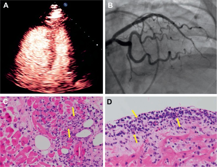
Figure 1.

(A) Contrast echocardiogram shows right ventricular dilation and reduced biventricular function. (B) Cardiac angiogram shows no obstructive lesion in the left coronary system. (C, D) Endomyocardial biopsy with hematoxylin & eosin stain shows infiltrating lymphocytes with associated myocyte injury. Endocardial infiltrate includes lymphocytes and eosinophils (yellow arrows).
