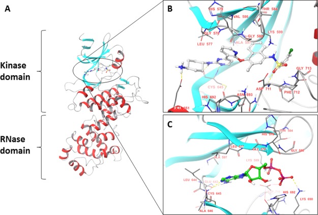
Figure 1.
(A) Ribbon diagram representing the structure of the IRE1 kinase and RNase domains (PDB code: 4U6R). β-strands are shown in blue and α-helices in red. Binding mode of (B) exogenous ligand kinase-inhibiting RNase attenuators (KIRAs) (PDB code: 4U6R) and (C) ADP (endogenous ligand) (PDB code: 3P23).

