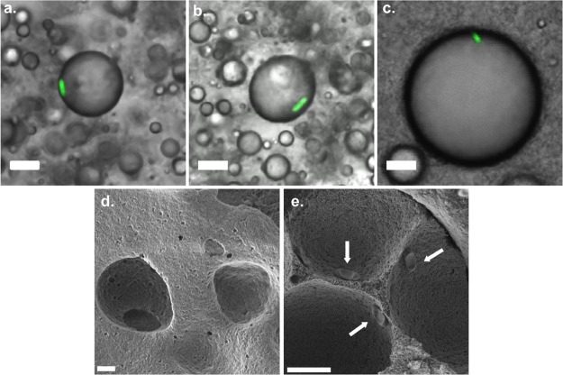
Figure 3.
Confocal microscopy images of single cell encapsulation of M. brunneumMa7-GFP conidia in a silica-NH2 Pickering emulsion (o/w ratio, 20:80) with different NPs contents of (a) 2, (b) 3 and (c) 5 wt %. Scale bar is 10 μm. SEM micrographs of dried silica-NH2 Pickering emulsion, (d) without conidia and (e) with conidia (arrows).

