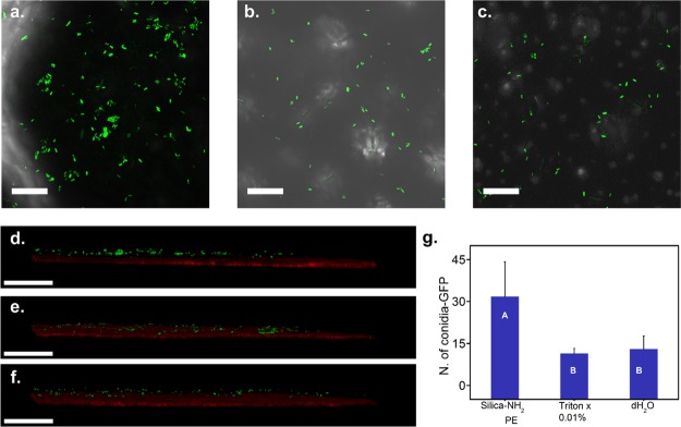
Figure 4.
Distribution of M. brunneum conidia-GFP on R. communis leaves. (a–c) Z projection and (d–f) cross section of confocal microscopy images of M. brunneum-GFP conidia after spray application on the surface of the leaves. (a,d) Conidia in silica-NH2 Pickering emulsion; (b,e) conidia in 0.01% Triton X-100 in water; (c,f) conidia in distilled water. (g) Number of conidia on leaves. Bars with the same letter do not differ significantly in the number of spores; Students t-test (P = 0.05). Scale bar: (a–c), 100 μm. (d–f), 200 μm.

