Figure 3.
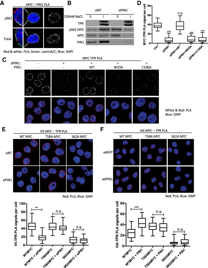
PIN1-mediated isomerization promotes pS62MYC association with the nuclear pore. (A) PLA of PIN1 and total MYC or pS62MYC in HeLa cells. The far left panels show the detailed colocalization of PIN1–MYC PLA with the nuclear envelope marker Lamin A/C. (B) Western blot of subcellular fractionation of pS62MYC and total MYC in nuclear-soluble (S) and nuclear-insoluble (I) fractions of HeLa cells transfected with nontargeting (NT) control or PIN1 targeted siRNAs and extracted by 150 nM NaCl. (C) PLA of MYC–TPR association in HeLa cells transfected with nontargeting or PIN1 siRNAs and the indicated PIN1 expression constructs. The expression of relevant proteins is shown in Supplemental Figure S3B. (D) Quantification of C showing box plots of PLA signals. The whiskers represent 5%–95% intervals, and the box represents median and 25%–75% intervals of PLA signals per cell from 50 cells per condition. Data are representative of three independent experiments. (E) PLA of V5-MYC–TPR association in HeLa cells transfected with nontargeting or PIN1 siRNAs and V5-tagged wild-type, T58A, and S62A MYC expression constructs. Quantification box plots are as in D. The expression of relevant proteins and grayscale images are shown in Supplemental Figure S3D and F, respectively. (F) PLA of HA-MYC–TPR association in primary MEF cells with Cre-dependent ROSA knock-in-expressed HA-tagged wild-type, T58A, and S62A MYC. Cells were coinfected with adenovirus Cre for MYC expression and GFP (adGFP) or PIN1 (adPIN1). Quantification box plots are as in D. The expression of relevant proteins and grayscale images were shown in Supplemental Figure S3E and G, respectively. P-values are shown for relevant significant comparisons. (**) P < 0.01; (***) P < 0.001.

