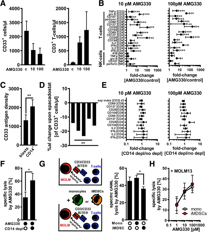
Fig. 2.
AMG 330 triggers T-cell-mediated lysis of AML-blasts that is further enhanced by MDSC depletion. (a) The absolute number of CD33+ AML-blasts and CD3+ T-cells was quantified in patient-derived AML PBMCs (n = 10) after 6 days of treatment with control BiTE® antibodies (c) or AMG 330. (b) AML-derived PBMCs (n = 12) were treated with control BiTE® antibodies or AMG 330 for three days. The median fluorescence intensity (MFI) of TNFα, granzyme B (grz B), CD107, perforin, CD69, CD137, CD25, CD154, IL2, and IFNγ was assessed by FACS in CD4+/CD8+ CD3+ T-cells and CD56+CD3neg NK-cells as indicated. The cells’ MFI from samples treated with control antibodies was set as 1. (c) CD33 surface antigen quantification was performed for AML-blasts and CD14+ monocytes (n = 8). (d) AML-derived PBMCs (n = 5) were treated with 10 pM AMG 330 in the presence or absence of the IDO inhibitor epacadostat (1 μM) and the number of CD33+ AML-blasts quantified. The graph displays the individual %al changes in cell numbers in presence of epacadostat. (e) AML PBMCs (n = 7) with/without prior depletion of CD14+ cells were treated with AMG 330 for three days. Expansion index and MFI of CD69, CD137, CD25, CD154, IL2, and IFNγ were assessed by FACS in VPD450-labeled CD4+/CD8+ CD3+ T-cells. Samples without depletion of CD14+ cells were set as 1. (f) AML-derived PBMCs (n = 5) with/without prior depletion of CD14+ cells were treated with AMG 330 for six days. LDH release as a surrogate for cell lysis was measured in the cultures’ supernatants. (g) Calcein-labeled MOLM-13 cells (MOLM) were co-cultured with T-cells alone (upper illustration) or with T-cells together with autologous monocytes or AML-educated iMDSCs (n = 5) +/− AMG 330 (lower illustration). Specific lysis of MOLM-13 cells was assessed after 3 h. (h) Calcein-labeled monocytes or iMDSCs (n = 4) were co-cultured with autologous T-cells and MOLM-13 cells +/− AMG 330. Specific lysis of monocytes/iMDSCs was assessed after 3 h. Bars indicate the standard error of the mean. Abbreviations: *, p < 0.05; **, p < 0.01; ***, p < 0.001

