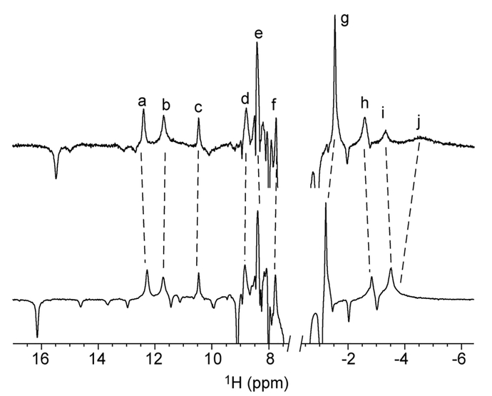
Figure 4.
The 1H NMR inversion recovery spectra of H46L mesoGlbN (top, 0.5 mM, 99% 2H2O, pH* 7.3) and H46L/Q47L mesoGlbN (bottom, 2 mM, 99% 2H2O, pH* 7.2) using a 50 ms recovery delay. Positive peaks identify protons within ~6 Å of the ferric iron. a, His70 Hβ3; b, His117 Hβ3; c, Phe84 Hζ; d, mesoheme δ-meso; e, Phe84 Hε1/ε2; f, His117 Hα; g, Val74 Hγ1; h, mesoheme a-meso H; i, mesoheme γ-meso H; j, unassigned.

