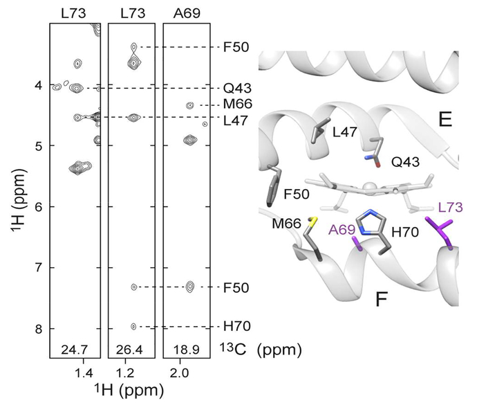
Figure 9.
NOESY cross peaks observed for Leu73 and Ala69 in H46L/Q47L mesoGlbN. Portions of the 13CH- selective NOESY data displaying the CH- groups of Leu73 and Ala69 are shown on the left. Cross peaks to Phe50 Hβ3, Gln43 Hα, Met66 Hα, Leu47 Hα, Phe50 Hδ1/δ2 and His70 Hα are labeled. Unlabeled peaks correspond to intraresidue NOEs. The relevant residues are shown on the right using the crystal structure of cyanomet GlbN (PDB ID: 1S69).14 Leu73 and Ala69 are colored purple. The Gln47Leu replacement was generated with Chimera.61 In this structure the distance between Leu7- and Leu47 is greater than 15 Å. The observed NOEs require the relative repositioning of the E and F helices and the removal of the intervening heme.

