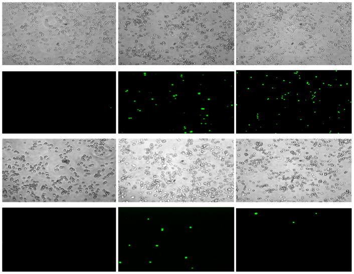
Figure 5.
Fluorescence microscopy (10× magnification) showing bright field (panels 1–3 and 7–9) and fluorescent images (panels 4–6 and 10–12) of macrophages in the presence of either 2 μM CM15 (panels 1–6) or 32 μM D3,7,13 (panels 7–12) immediately after peptide addition (column 1), and after incubation at 37°C for 15min (column 2), or 30min (column 3).

